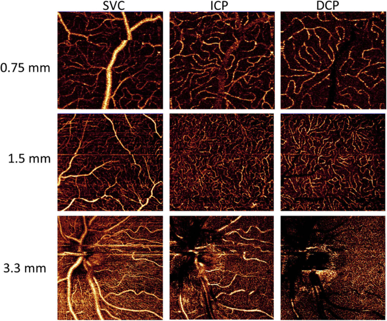
Fig. 8.
OCTA of FOV of 0.75×0.75 mm at 5 degrees superior to the fovea, 1.5×1.5 mm at 5 degrees nasal to the fovea and 3.3×3.3 mm of the peripapillary area, acquired from an eye with −0.5 diopters of defocus and 0.25 diopters of astigmatism. High capillary contrast is observed for the superficial vascular complex (SVC), intermediate capillary plexus (ICP) and deep capillary plexus (DCP). The low prevalence of OCTA projection artifacts is visualized on ICP and DCP images.

