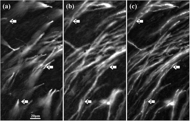
Fig. 7.
Imaging results of brain tissue: typical raw images are captured with the conventional system (a) and the proposed system (b). (c) The Maximum-Intensity projection along the propagation axis with the conventional system. Arrows 1, 2 and 3 represent structures in the z plane of -20 , 0 , 10 .

