. 2020 Sep 23;128(9):097006. doi: 10.1289/EHP6470

Figure 3.

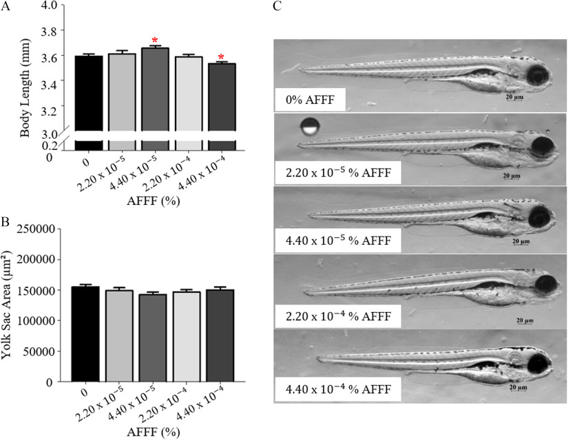
Figures 3A and 3B are bar graphs, plotting body length (millimeter) and Yolk Sac Area (micrometer squared), ranging from 0 to 0.2 in increments of 0.2, 0.2 to 3.0 in increments of 2.5, and 3.0 to 4.0 in increments of 0.2 and 0 to 250000 in increments of 50000, respectively, (y-axis) across aqueous film-forming (percent), including 0, 2.20 times 10 begin superscript negative 5 end superscript, 4.40 times 10 begin superscript negative 5 end superscript, 2.20 times 10 begin superscript negative 4 end superscript, and 4.40 times 10 begin superscript negative 4 end superscript (x-axis). Figure 3C is a schematic diagram of five different treatment groups for larval development with aqueous film-forming exposures, including 0 percent aqueous film-forming, 2.20 times 10 begin superscript negative 5 end superscript percent aqueous film-forming, 4.40 times 10 begin superscript negative 5 end superscript percent aqueous film-forming, 2.20 times 10 begin superscript negative 4 end superscript percent aqueous film-forming, and 4.40 times 10 begin superscript negative 4 end superscript percent aqueous film-forming.

Larval developmental measurements at 96 h post fertilization (hpf) with AFFF exposures. Larval body length (A) and yolk sac area (B) are graphed. (C) Representative images from 04.40×10⁴% AFFF treatment groups. Bars represent mean±SEM. Asterisk (*) indicate p<0.05 compared with 0% AFFF, one-way ANOVA, Tukey’s HSD post hoc test. n=5456 larvae per treatment group. Note: AFFF, aqueous film-forming foam; ANOVA, analysis of variance.