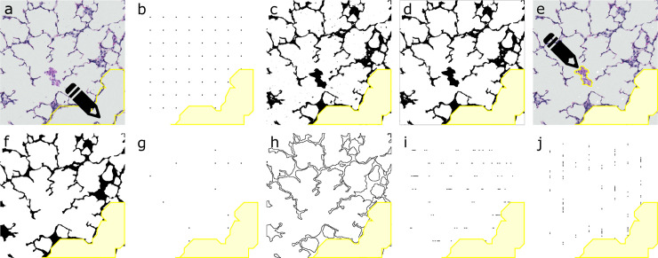
Fig 1. Summary of image processing and analysis from lung field to Pref, Psep and I*.
(A) Non-parenchymal tissue (in this case conductive airway) is selected manually. (B) Pref is counted as the number of points of a 64 point test grid, outside the selection of non-parenchymal tissue. (C) Image is thresholded to binary mask. (D) Automatically all small and unconnected exudates are removed. (E) Larger and connected exudates require manual selection. (F) Mask is cleaned and smoothened. (G) Psep is counted as the number of points of the test grid that are common to the cleaned mask. (H) The edges of the cleaned mask are aligned. (I-J) I is counted as the number of intersections of a test system of 8 horizontal and 8 vertical lines with the edge-image. *Pref = reference points, Psep = septal points, I = intersections.

