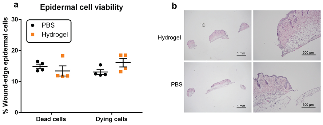
Figure 5.

Hydrogel effects on cells in the wound environment. (a) Percent of dead and dying epidermal cells at the wound edge in hydrogel H2-treated and PBS buffer-treated mice. (b) Hematoxylin and eosin staining of tissue 24 hours after hydrogel and buffer treatment; no significant differences were noted.
