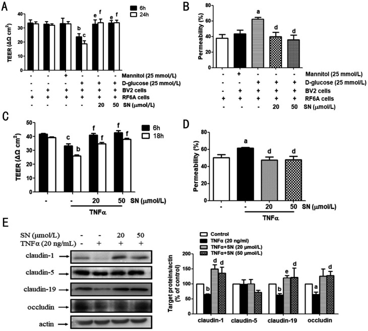
Figure 4. SN rescued the D-glucose-stimulated BV2 cells or TNFα-induced iBRB injury in RF/6A cells.
A, B: Upper-chamber seeded with RF/6A cells and lower-chamber seeded with BV2 cells in 24-well plates composed a co-cultured environment, and 6h before the D-glucose (25 mmol/L) stimulation on BV2 cells, SN (20, 50 µmol/L) was added into the bottom of transwells for pre-treatment. TEER (n=3) and FITC-dextran leakage (n=4) were determined. C, D: TNFα (20 ng/mL) was added into the plates under the chambers, and RF/6A cells were cultured in the upper-chamber with the pretreatment with or without SN (20, 50 mmol/L) for 6h. TEER (n=3) and FITC-dextran leakage (n=4) were detected. E: The same treatment as C&D was performed and the cell samples were collected for detecting the content of claudin-1 (n=4), claudin-5 (n=4), claudin-19 (n=3) and occludin (n=4) aP<0.05, bP<0.01, cP<0.001 versus control; dP<0.05, eP<0.01, fP<0.001 versus D-glucose-treated BV2 cells or TNFα.

