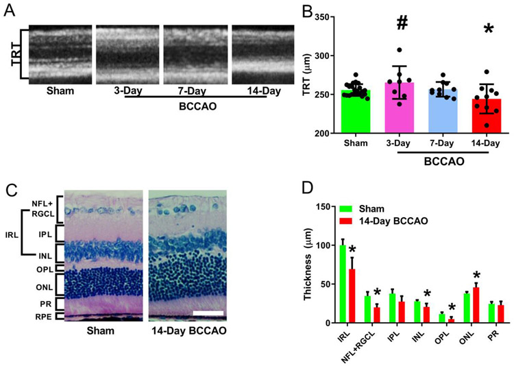
Figure 5. Morphological changes and thickness measurements in the retinal layers following long-term BCCAO.
(A) Representative optical coherence tomography images of the retina. (B) The resulting images were analyzed using the Heidelberg Spectralis system software to measure the total thickness. (C) Example Hematoxylin and Eosin (H&E) sections were used to compare layer thickness (original magnification 400X). (D) H&E thickness quantification of each layer in different groups. The data were presented as mean ± SD. *p<0.05 vs Sham. Layer name abbreviations: NFL: nerve fiber layer; RGCL: retinal ganglion cell layer; IPL: inner plexiform layer; INL: inner nuclear layer; IRL: inner retinal layer; OPL: outer plexiform layer; ONL: outer nuclear layer; PR: photoreceptor layer; RPE: retinal pigment epithelium. Scale bar= 50 μm.

