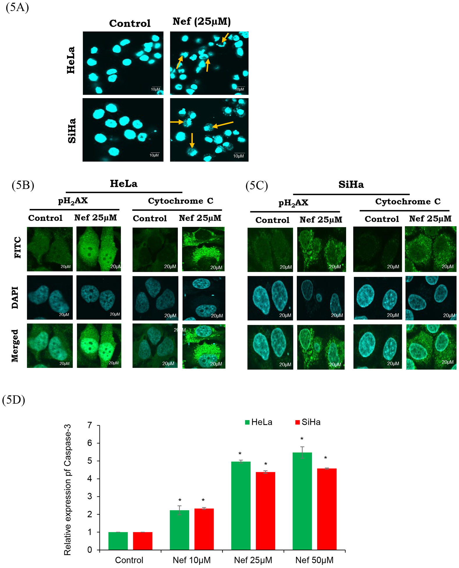
Figure 5: Nef induces apoptosis by activating apoptotic characteristics:

(A) Nuclear morphology of HeLa and SiHa cells stained with DAPI. Apoptotic bodies (arrows), cell swelling, and chromatin lysis were observed. Fluorescence microscopy images are of 60× magnification with scale bar of 10μm. HeLa (B) and SiHa (C) cells treated with Nef (25μM) were fixed, blocked and probed with pH2AX and cytochrome-C antibodies. Then the cells were probed with secondary antibody conjugated with FITC (green) and counter stained with DAPI (blue). Nef treated cells showed activation of pH2AX and cytochrome c compared to untreated control cells. (D) Quantitative analysis of relative expression of caspase-3 by ELISA. Both HeLa and SiHa cells treated with Nef show significant increase in the relative expression of caspase-3 in comparison to untreated control cells. Results are representative of 3 independent experiments. *P<0.01 when compared with the control group.
