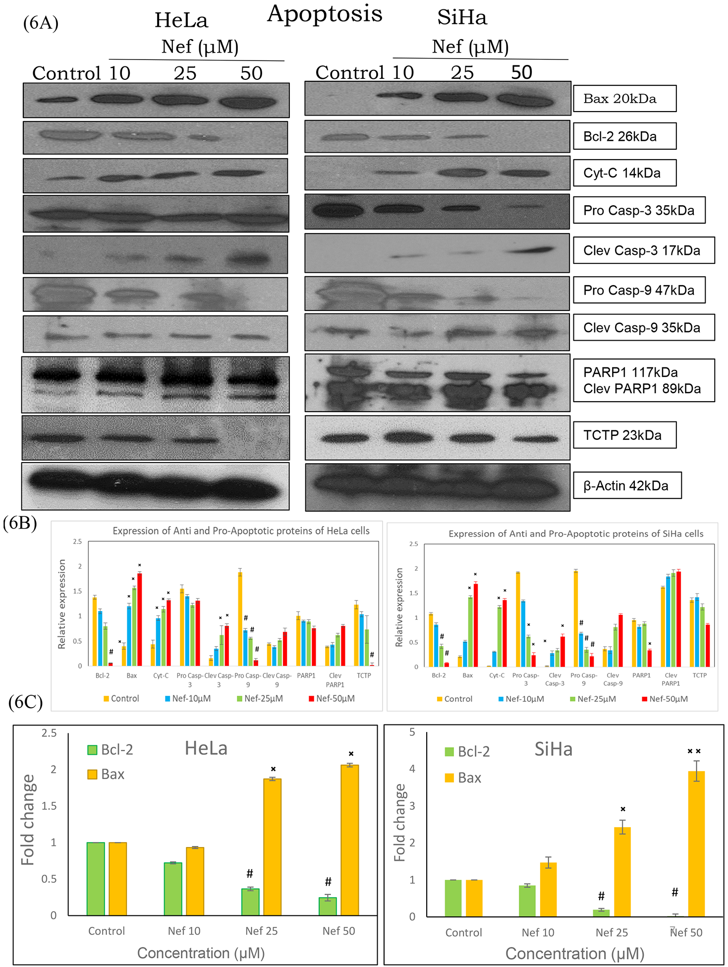
Figure 6:

(A) Western blot and real time-PCR analysis of apoptosis markers in HeLa and SiHa cells. Cell lysates of HeLa and SiHa cells treated with Nef (10, 25 and 50μM) for 48hrs were subjected to SDS-PAGE, transferred to nitrocellulose membrane and probed with respective primary antibodies. Results revealed that Nef treatment increased the expression of pro-apoptotic markers bax, cytochrome c, cleaved caspase-3, −9 and PARP while reducing the levels of anti-apoptotic bcl-2, TCTP, procaspase-3 and −9. Image shown is the chemiluminescent detection of the blots. (B) Relative expression of the proteins were normalized using β-actin as loading control and band intensities were quantified using Image J software. *p < 0.01 or #p<0.01, as compared with the control. (C) mRNA expression of bcl-2 and bax was determined by real time-PCR. Results revealed that Nef upregulated the expression of Bax with concurrent downregulation of Bcl-2. β-actin served as house keeping control in this experiment. Data are representation of one of three experiments. *p < 0.01 or #p<0.01, as compared with the control.
