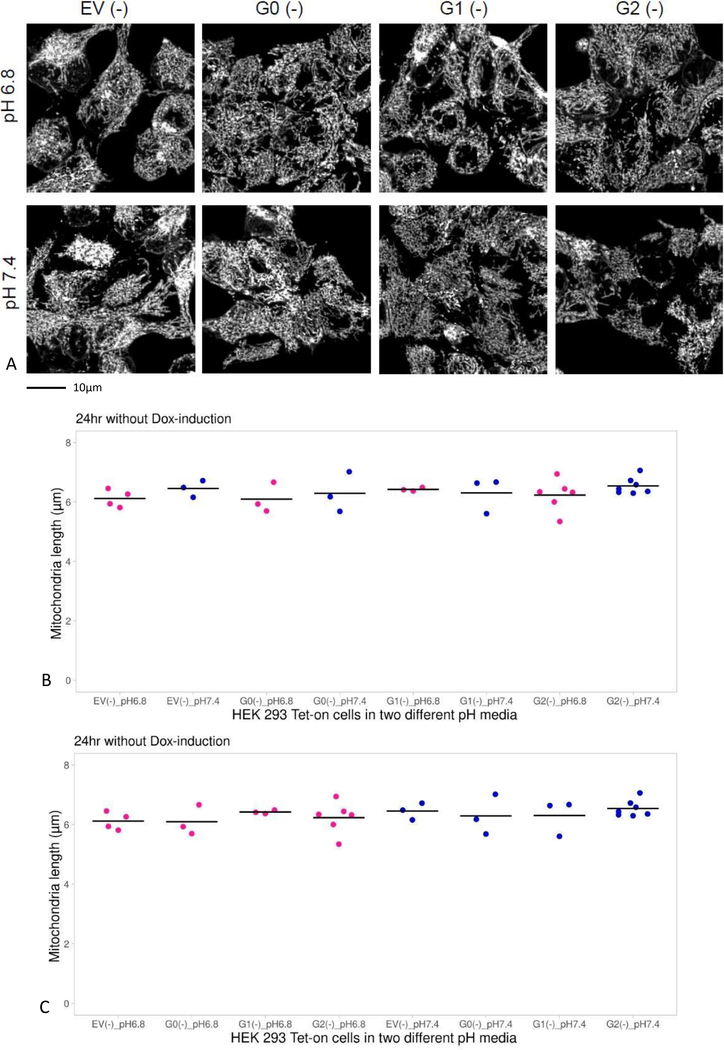
Figure 2. Comparison of mitochondrial length in HEK293 Tet-on cells cultured in different media pH without doxycycline (Dox).
Panel A. Images displaying mitochondrial morphology in cells cultured in growth media pH 6.8 or 7.4 without Dox (absence of APOL1 induction). The cells were labeled with MitoTracker Red, a red-fluorescent dye that labels mitochondria in live cells and imaged on laser scanning confocal microscope with Olympus super-apochromat oil objective at 1000x magnification. The cells were 3D rendered and processed using Fiji software (a derivative of Image J) and MiNA plug in. The cells were kept in a sealed, environmentally controlled chamber gassed with 5% CO2 at temperature 37°C during imaging, so that stable pH of 7.4 or 6.8 was maintained in the same manner as in an incubator.
Panel B. Graphs display a summary of the measurements of mitochondrial length in HEK293 Tet-on cells over 24 hours when APOL1 was not induced. In the absence of Dox, mitochondrial length was comparable in HEK293 cells irrespective of media pH or APOL1 genotype (p=NS, ANOVA); results grouped according to genotype.
Panel C. Same as Panel B, except that data grouped according to media pH (p=NS, ANOVA).

