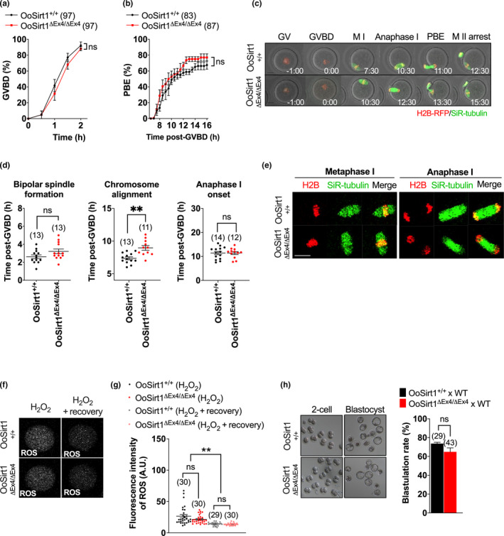
FIGURE 2.

In vitro maturation, spindle assembly, chromosome alignment and segregation in oocytes and preimplantation development of embryos from young OoSirt1+/+ and OoSirt1ΔEx4/ΔEx4 females. Rates of (a) GVBD and (b) PBE. (c) Shown are panels comprised of selected brightfield and fluorescence frames from representative timelapse series of OoSirt1ΔEx4/ΔEx4 and OoSirt1+/+ oocytes. Time, h:min relative to GVBD. (d) Quantification of timing of bipolar spindle formation, chromosome alignment and anaphase I‐onset. (e) Shown are representative images of live OoSirt1ΔEx4/ΔEx4 and OoSirt1+/+ oocytes during metaphase I and anaphase I. (f) Shown are representative images of ROS fluorescence in oocytes immediately following peroxide treatment and after 90 min of recovery. (g) ROS quantification in peroxide treated OoSirt1ΔEx4/ΔEx4 and OoSirt1+/+ oocytes. (h) Blastulation rates of embryos derived from OoSirt1ΔEx4/ΔEx4 and OoSirt1+/+ females crossed with WT males. Shown are representative brightfield images of 2‐cell and blastocyst stage embryos. Oocyte and embryo numbers are shown in parentheses. Scale bar = 20 µm. Data are shown as mean ± SEM. Statistical analyses performed using two‐way Anova with Sidak's multiple comparisons test (a, b) or two‐tailed Student's t test (d, g, h). p values are represented as **p ≤ 0.01, ns denotes p > 0.05
