
FIGURE 2.
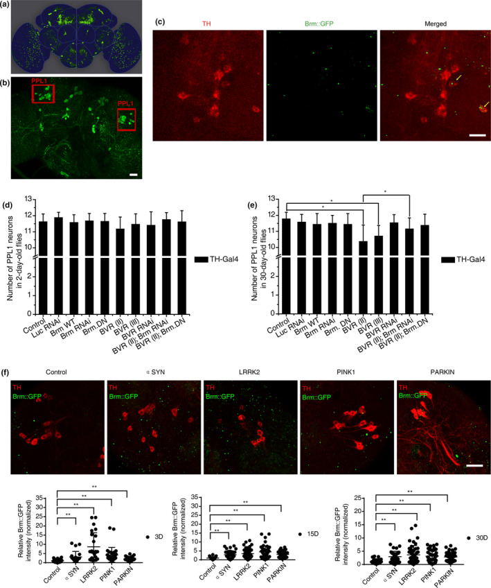
Inhibition of Brahma rescued dopaminergic (DA) degeneration caused by overexpression of BVR in Drosophila. (a) Schematic diagram of representative DA neuronal clusters in adult Drosophila brain. (b) Representative images show the posterior DA clusters in the fly brain. The intact left or right PPL cluster contains ~12 DA neurons at average in healthy adult flies. The DA neurons were labeled with green fluorescent protein (GFP). Scale bars, 25 μm. (c) Expression of Brm in the Drosophila DA neurons. A fusion Brm::GFP transgene was used to report the endogenous protein expression level of Brm. The Brm protein (green) was present in the Drosophila PPL1 DA neurons (anti‐TH, red) (pointed with yellow arrows) but not limited in DA neurons. Scale bars, 10 μm. (d, e) Scoring PPL1 DA neurons in 2‐day‐old (d) and 30‐day‐old (e) flies subjected to Brm‐ or BVR‐related genetic manipulations. Genetic manipulations included TH‐Gal4 driving overexpression of wide type Brm (Brm wt), a dominant‐negative allele of Brm (BrmDN), dBVR OE (Bvr II and Bvr III), Bvr II + BrmDN and induction of Brm RNAi, BVR RNAi or Bvr II +Brm RNAi with w‐ (TH‐Gal4/+) and Luc RNAi (TH‐Gal4/UAS‐Luc RNAi) flies as the control. The genotypes of experimental flies are provided in the Appendix S2. Overexpression of dBVR resulted in DA neuronal loss in the aged fly brains when compared with controls (Bvr II, 10.37 ± 1.03; Bvr III, 10.70 ± 0.67; and control, 11.77 ± 0.42). n > 20 for each data point. (f) Progressive elevation of Brm protein levels (GFP signal) in the brains of four PD model flies in comparison to the control. Representative whole‐mount fluorescence images of fly brains are provided, with quantifications of the Brm protein level in young (3‐day‐old), middle‐age (15‐day‐old), and aged (30‐day‐old) flies shown below (n > 5). *indicates Mann–Whitney p < 0.01. Scale bar, 10 μm