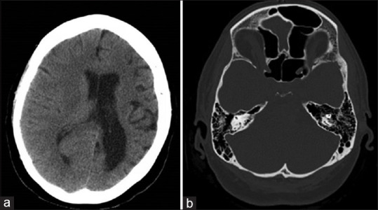
Figure 1.

(a) Head computed tomography scan 2018 showing hemiatrophy of the left cerebral hemisphere with volume loss, ex-vacuo dilatation of the body, frontal horn, temporal horn, and occipital horn of the left lateral ventricle. (b) Head computed tomography showing sinus hyperaeration
