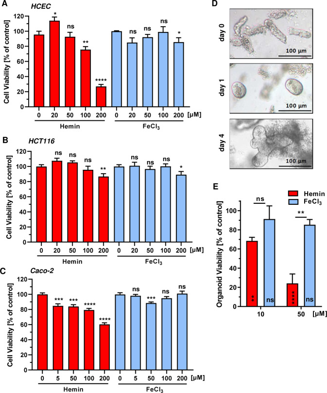
Fig. 2. Impact of heme iron and inorganic iron on cell and intestinal organoid viability.
a–c HCEC (a), HCT116 (b) and Caco-2 (c) were treated with increasing concentrations of hemin or FeCl3 (0–200 µM). Cell viability was determined after 72 h using the MTS assay. Data are given as mean + SEM (n ≥ 3, triplicates). Ns: p > 0.05; *p < 0.05; **p < 0.01; ***p < 0.001; ****p < 0.0001 (versus respective control). d Microscopic images of isolated intestinal crypts (day 0) and the developing intestinal organoids (day 1 and 4). e Intestinal organoids were treated with hemin or FeCl3 for 24 h and viability was determined by the MTT assay. Date are given as mean + SEM (n = 2). Ns: p > 0.05; **p < 0.01; ****p < 0.0001.

