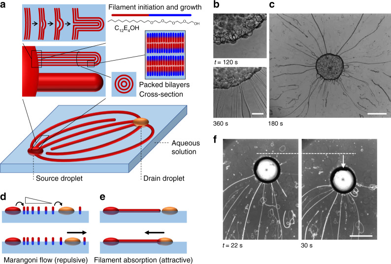
Fig. 1. Marangoni flows and filaments direct positioning.
a In the amphiphile source droplet (red sphere), the C12E4OH amphiphile forms closely packed bilayers. At the boundary of the droplet that is floating on water, the amphiphiles take up water (light blue), the packed bilayers buckle, and form long filaments consisting of bilayers. b, c Optical microscopy images of filament extrusion from the amphiphile source droplet, after deposition at an aqueous sodium alginate solution at t = 0 s. The scale bar in b represents 200 μm; the scale bar in c is 1 mm. d Concomitant to the filament growth, individual amphiphile molecules are released from the source droplet (red sphere) to the air–water interface. Subsequently, these amphiphiles are depleted at the drain droplet (orange sphere), such that a Marangoni flow emerges that pushes the source and drain droplets apart. e The drain, however, is pulled back to the source upon absorption of the filaments. A dynamic organization emerges when the repulsive (Marangoni flow) and attractive forces (filament absorption) match. f Upon filament absorption, the drain droplet (deposited at t = 0 s) is pulled towards the source—positioned in the bottom with respect to the area that is featured in the microscopy images. The scale bar represents 1 mm.

