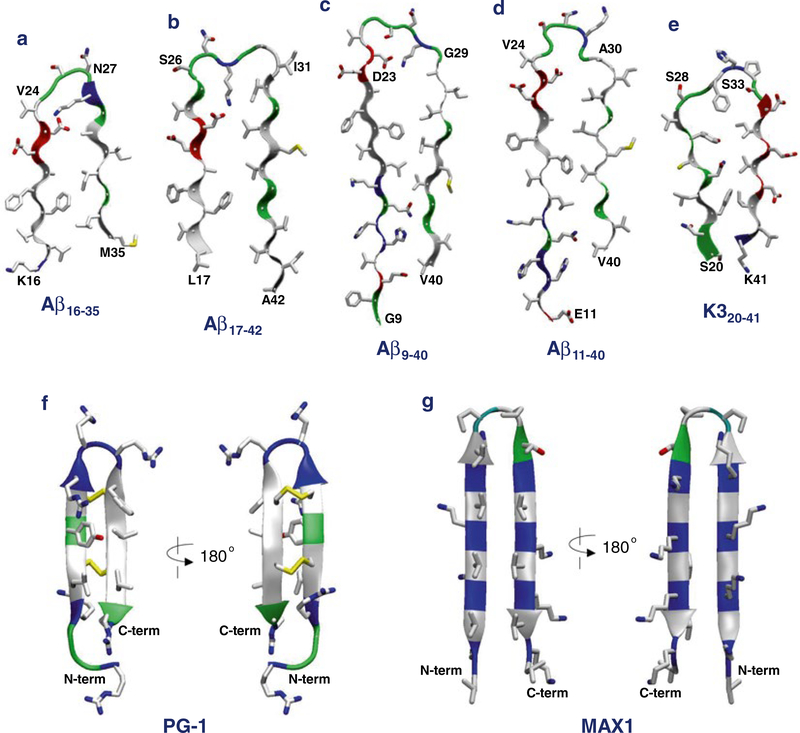
Fig. 1.
Monomer conformations recruited for the molecular dynamics (MD) simulations of β-sheet channels in the lipid bilayers. The U-shaped amyloid peptides with the β-strand-turn-β-strand motif for (a) the computational Aβ16–35, (b) the NMR-derived Aβ17–42, (c) the ssNMR Aβ9–40, (d) the ssNMR Aβ11–40, and (e) the ssNMR K320–41 peptides. The β-hairpin motif for the synthetic (f) protegrin-1 (PG-1) and (g) MAX1 peptides. In the peptide ribbon, hydrophobic, polar/Gly, positively charged, and negatively charged residues are colored white, green, blue, and red, respectively. Yellow sticks in PG-1 denote the disulfide bonds

