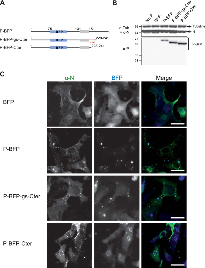
FIG 4.
Study of the role of the intrinsically disordered domain [160-227] of P for IB formation. (A) Schematic illustration of the full-length and truncated P-BFP proteins. The oligomerization domain of P is represented in gray. The BFP is shown in blue, and the sequence of five Gly-Ser repetition is shown in red. Numbers indicate amino acid positions. (B) Western blot analysis of the expression of N and P-BFP proteins in BRST/7 cells cotransfected with pN- and pP-BFP-derived constructs. Detection of tubulin was used as a control. (C) Cellular localization of N and P-BFP proteins in cells. N and P-BFP proteins were coexpressed in BSRT7/5 cells. The cells were then fixed 24h posttransfection and labeled with anti-N (green) antibodies, and the distribution of viral proteins was observed by fluorescence microscopy. Bars, 20 μm.

