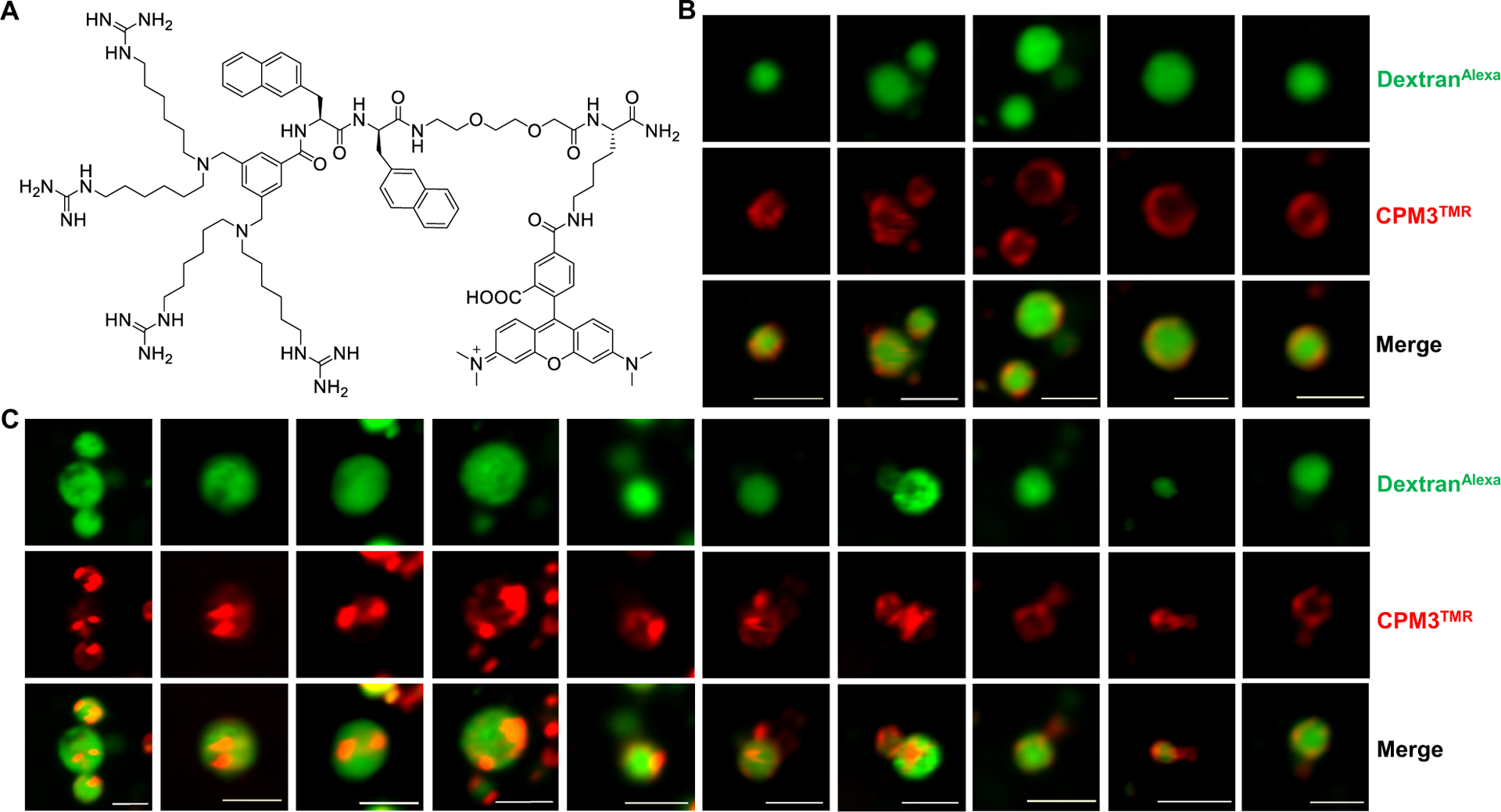
Figure 5.

CPM3-induced vesicle budding and collapse from enlarged endosomes. (A) Structure of CPM3TMR. (B,C) Live-cell confocal microscopic images of endosomes at different stages of vesicle budding and collapse process. HeLa cells were pretreated with YM201636 (800 nM) for 2 h, and CPM3TMR (2 μM, red channel) and DextranAlexa (50 μg/mL, green channel) were added. After incubation for another 30–40 min, the cells were washed and imaged by live-cell confocal microscopy. The images shown are snapshots of different endosomes from multiple cells. Scale bars = 2 μm.
