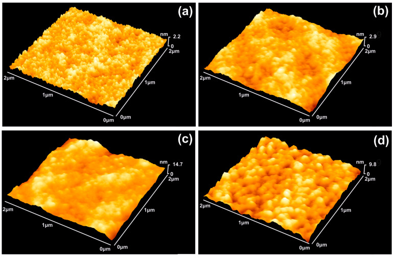
Figure 1.
AFM surface imaging of PHEMA layers. The scan area is 2 × 2 μm2 and the 157 nm laser fluence 250 Jm−2 per pulse: (a) Non-irradiated PHEMA layer; (b) Irradiation with 20 laser pulses (lp), 5 kJm−2; (c) 70 lp, 17.5 kJm−2; (d) 200 lp, 50 kJm−2. The structure of the surface is constantly modified under laser irradiation up to 200 lp.

