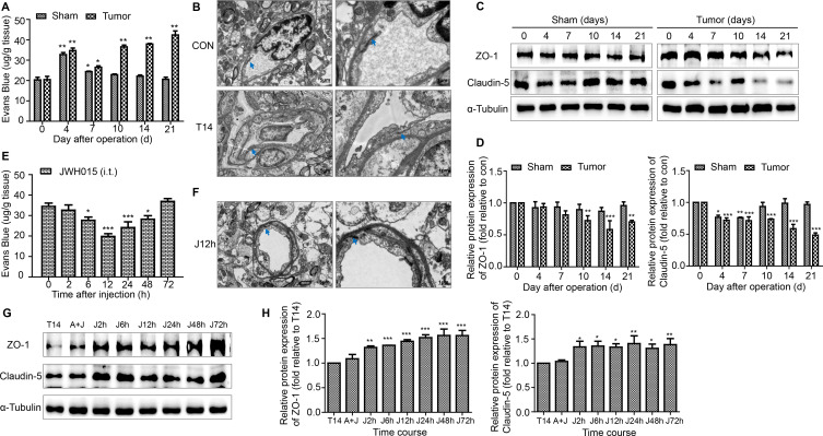
Figure 2.
Disruption of the BSCB in BCP mice. (A) The EB leakage in the lumbar enlargement of spinal cord were tested on days 0, 4, 7, 10, 14 and 21 after operation in the sham and tumor groups. n=3 for each time point per group. (B) Representative images of the ultrastructure of the BSCB in the lumbar enlargement obtained using transmission electron microscopy on day 0 and day 14 in tumor group. Scale bar, 5 µm and 1 µm. n=3 per group. (C, D) Representative blots and quantification of TJ proteins ZO-1 and claudin-5 in the spinal cord on postoperative days in the sham and tumor groups. n=3 for each time point per group. *P<0.05 vs day 0, **p<0.01, ***p<0.001. Treatment of JWH015 improved the disruption of BSCB in BCP mice. (E) The EB leakage in the lumbar enlargement of spinal cord were tested before the injection (0 hour) and at 2, 6, 12, 24, 48 and 72 hours after the injection of JWH015 in the tumor group. n=3 for each time point per group. (F) Representative images of the ultrastructure of the BSCB in the lumbar spinal cord, which was taken from 12 hours after the injection of JWH015, obtained using transmission electron microscopy. Scale bar, 5 µm and 1 µm. n=3. (G, H) Representative blots and quantification of ZO-1 and claudin-5 at 2, 6, 12, 24, 48, and 72 hours after the injection of JWH015 compared with vehicle injection, respectively. n=3 for each time point per group. *P<0.05 vs vehicle injection, **p<0.01, ***p<0.001. All data are presented as the means±SD. Arrows showed TJ structures between the endothelial cells. BCP, bone cancer pain; BSCB, blood–spinal cord barrier; TJ, tight junctions.

