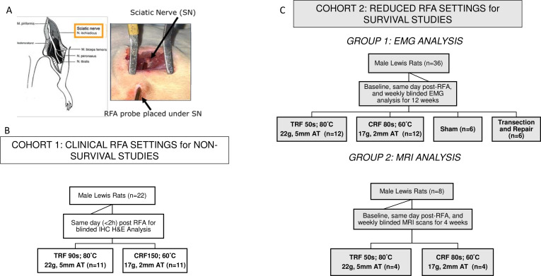
Figure 1.
Probe placement and Study Design. (A) The probe tip was placed directly under the sciatic nerve (parallel placement for TRF and angle independent for CRF) using visual guidance. (B) Cohort 1 included rats that were distributed into two treatment groups for same-day immunohistochemistry (IHC) analysis, using clinical run parameters (C) Cohort 2 included a group of rats that were survived for 12 weeks for EMG analysis and another group that was survived for 4 weeks for MRI analysis. CRF, cooled radiofrequency; EMG, electromyography; RFA, radiofrequency ablation; TRF, traditional radiofrequency.

