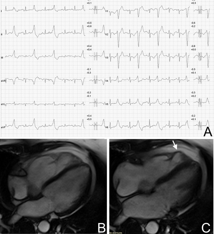
Figure 4.
Premature ventricular beats with a left bundle branch block-like pattern of the ectopic QRS and underlying right ventricular myocardial disease. Premature ventricular beats with a left bundle branch block/intermediate axis pattern, associated with ECG abnormalities (low QRS voltages in the limb leads and negative T-waves in V1–V3 in non-ectopic beats), which increased during exercise testing in a 34-year-old female runner (A). Cine cardiac magnetic resonance sequences (four-chamber view) revealed right ventricular dilation with hypertrabeculation (diastolic frame) (B) and dyskinesia (arrow) of the right ventricular free wall (systole frame) consistent with arrhythmogenic cardiomyopathy (C).

