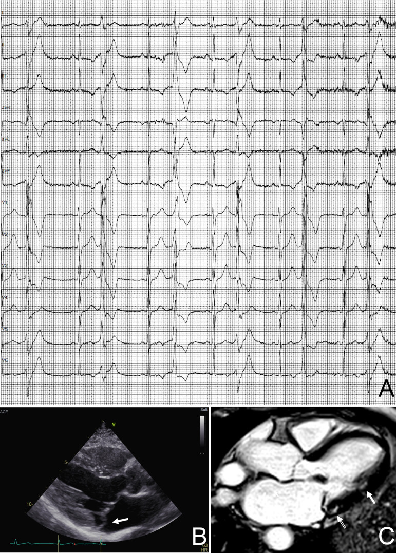
Figure 5.
Premature ventricular beats in an athlete with arrhythmic mitral valve prolapse. Premature ventricular beats with a right bundle branch block morphology and variable QRS axis, suggesting multiple ectopic foci in the left ventricular myocardium (A). Echocardiography (long-axis parasternal view) showing thickened and prolapsing mitral valve leaflets (arrow) (B). Postcontrast cardiac magnetic resonance sequence (apical four-chamber view) showing potentially arrhythmogenic areas of myocardial fibrosis/late gadolinium enhancement which are localised behind the posterior leaflet of the mitral valve (open arrow) and at the implant of the posterolateral papillary muscle (arrow) (C).

