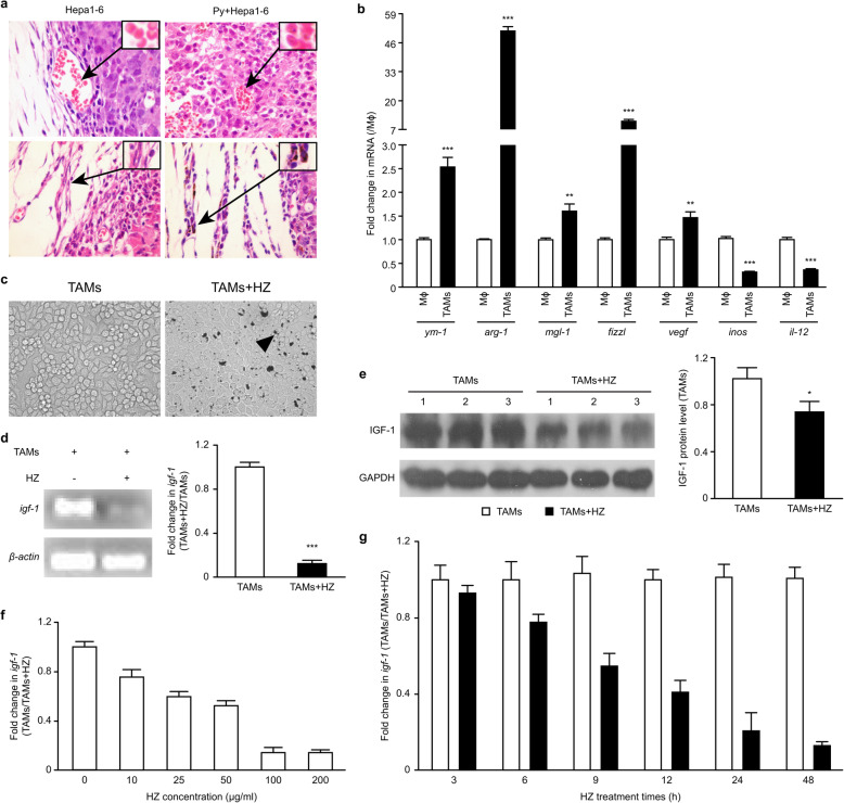
Fig. 6.
HZ modulated the expression of IGF-1 in TAMs polarized in vitro by coculture. a Tumor tissue sections were observed on day 17 after Plasmodium infection (n = 4). Parasite-infected red blood cells (iRBCs) could be found in the tumor tissue (upper panel). Representative photos are presented (1000× magnification). The insets show representative iRBCs, and the black arrows identify Plasmodium parasites in the iRBCs. HZ was found in TAMs from Hepa1–6 cell-implanted mice infected with Plasmodium parasites (n = 4) (bottom panel). The arrows indicate representative cells with HZ accumulation in their cytoplasm. Original magnification: 400×. RAW264.7 cells were plated in the absence or presence of TSN (1:2 dilution). b-c Genes associated with TAM phenotype characteristics were analyzed in the induced TAMs by real-time PCR (b). The phagocytosis of HZ by RAW264.7 cells in vitro was also observed (c). The arrows illustrate examples of cells with HZ accumulation in their cytoplasm. Original magnification: 400×. d-e The cells were treated with or without HZ (100 μg/ml) in the presence of TSN (1:2 dilution). The level of IGF-1 was analyzed 48 h after coculture by RT-PCR (d, left panel) and qRT-PCR (d, right panel). The expression of IGF-1 was detected by immunoblotting and quantified using ImageJ software (e). f-g In some experiments, the induced TAMs were treated with different doses of HZ, and 48 h later, the expression of IGF-1 mRNA was analyzed by qRT-PCR (f). In other experiments, the induced TAMs were exposed to HZ (100 μg/ml) for various time periods, and the level of IGF-1 was then analyzed (g). The results are presented as the means ± SDs from triplicate samples in three independent experiments

