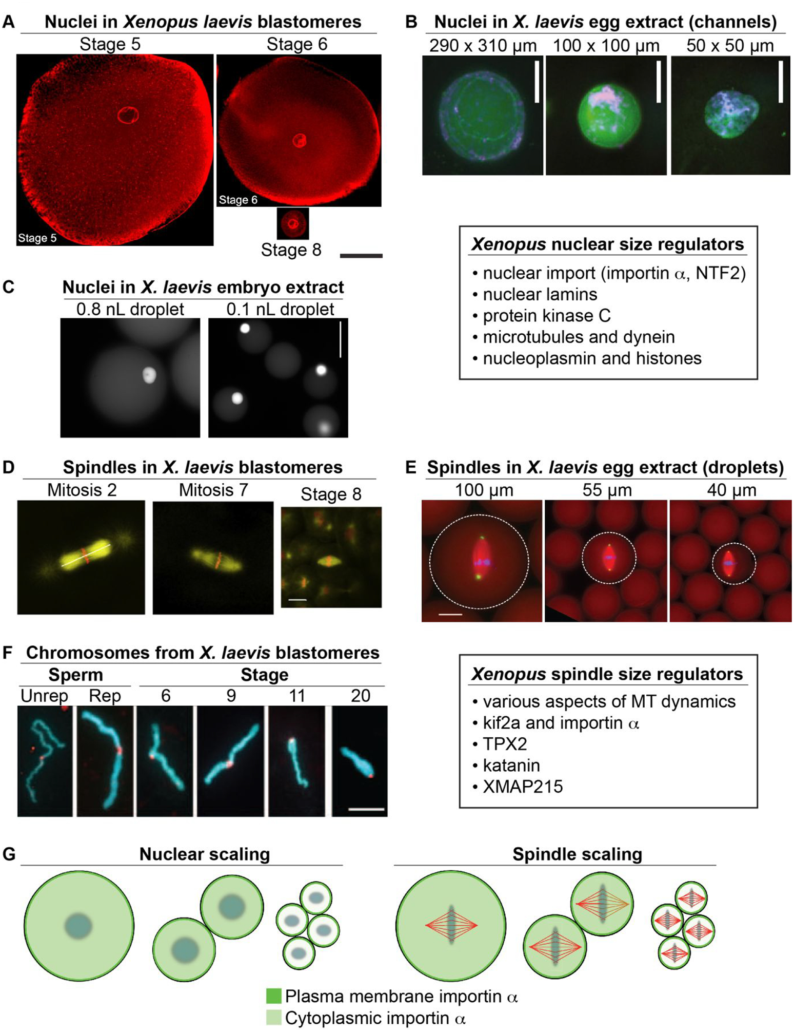
Figure 1: Developmental size regulation of intracellular structures in Xenopus.

(A) Blastomeres were isolated from different stage X. laevis embryos and stained for the NPC. Scale bar, 50 μm. Adapted with permission from (Jevtic & Levy, 2015). (B) Nuclei were assembled in X. laevis egg extract in microfluidic channels of varying dimensions. Channel dimensions are indicated as height × width. Membranes, DNA, and incorporated dUTP are labeled green, blue, and red, respectively. Scale bar, 20 μm. Adapted with permission from (Hara & Merten, 2015). (C) Nuclei and cytoplasm from stage 10 X. laevis embryos were encapsulated in droplets of varying volume and allowed to reach steady-state sizes. Droplet volumes are indicated above each panel. Nuclei are visualized by import of GFP-NLS. Scale bar, 50 μm. Adapted with permission from (P. Chen et al., 2019). (D) Different stage X. laevis embryos were fixed and stained for tubulin (yellow) and DNA (red). Scale bar, 20 μm. Adapted with permission from (Wuhr et al., 2008). (E) Spindles were assembled in different volumes of X. laevis egg extract. Tubulin, DNA, and NuMA are labeled red, blue, and green, respectively. Droplet diameters are indicated above each panel. Scale bar, 25 μm. Adapted with permission from (Hazel et al., 2013). (F) Condensed mitotic chromosomes from different stage X. laevis embryos are shown. For comparison, unreplicated (Unrep) and replicated (Rep) sperm chromosomes were incubated in X. laevis egg extract. DNA and kinetochores are labeled blue and red, respectively. Scale bar, 5 μm. Adapted with permission from (Kieserman & Heald, 2011). (G) As cells become smaller during early Xenopus development, palmitoylated importin α is increasingly partitioned to the plasma membrane. In the case of the nucleus, this reduces nuclear import and size. In the case of the spindle, reduced kif2a inhibition promotes MT depolymerization and spindle shortening. Adapted with permission from (Brownlee & Heald, 2019).
