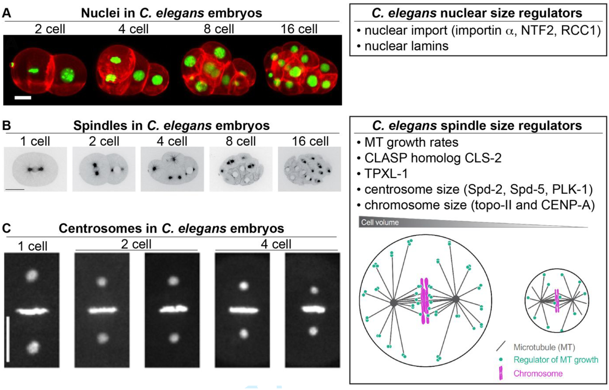
Figure 3: Developmental size regulation of intracellular structures in C. elegans.

(A) Different stage C. elegans embryos with nuclei labeled green for H2B and plasma membrane labeled red. Scale bar, 10 μm. Images adapted from (Fickentscher & Weiss, 2017) and made available under a Creative Commons Attribution 4.0 International License. (B) Different stage C. elegans embryos expressing GFP-tagged β-tubulin. Scale bar, 20 μm. Adapted with permission from (Lacroix et al., 2018). (C) Different stage C. elegans embryos expressing GFP-tagged γ-tubulin and H2B. Scale bar, 10 μm. Adapted with permission from (Greenan et al., 2010). The cartoon model in the “C. elegans spindle size regulators box” was adapted with permission from (Lacroix et al., 2018).
