
Figure 4: Developmental size regulation of intracellular structures in other model organisms.
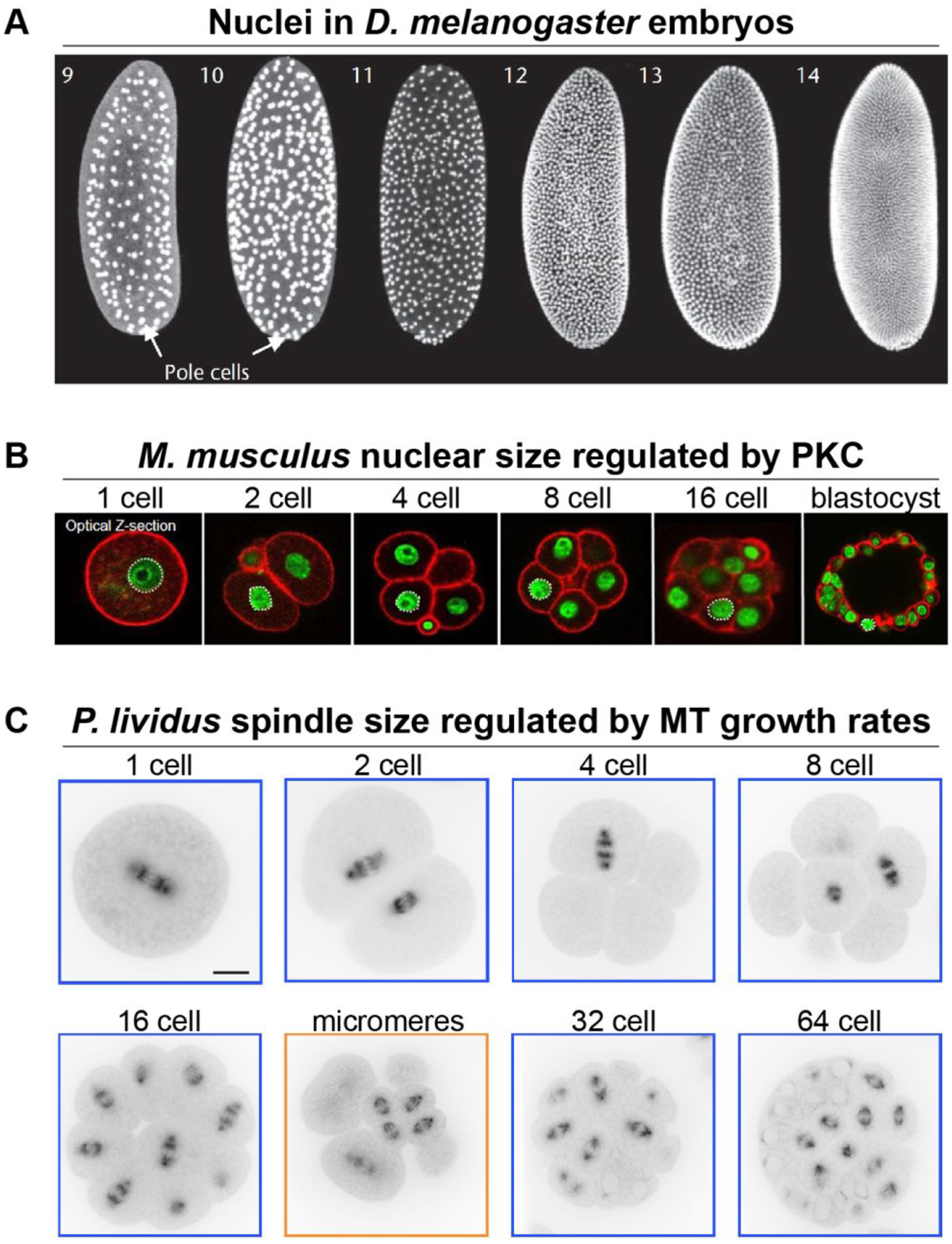
(A) Different stage D. melanogaster embryos showing labeled nuclei. Numbers denote the nuclear cycle. Note that nuclei are present in a syncytium until cellularization at nuclear cycle 14. Images adapted with permission from (Kotadia et al., 2010). (B) Different stage M. musculus embryos with DNA labeled green and cell cortex labeled red with phalloidin. Images adapted from (Tsichlaki & FitzHarris, 2016) and made available under a Creative Commons Attribution 4.0 International License. (C) Different stage P. lividus sea urchin embryos microinjected with ATTO 565-labelled tubulin. Scale bar, 20 μm. Adapted with permission from (Lacroix et al., 2018).