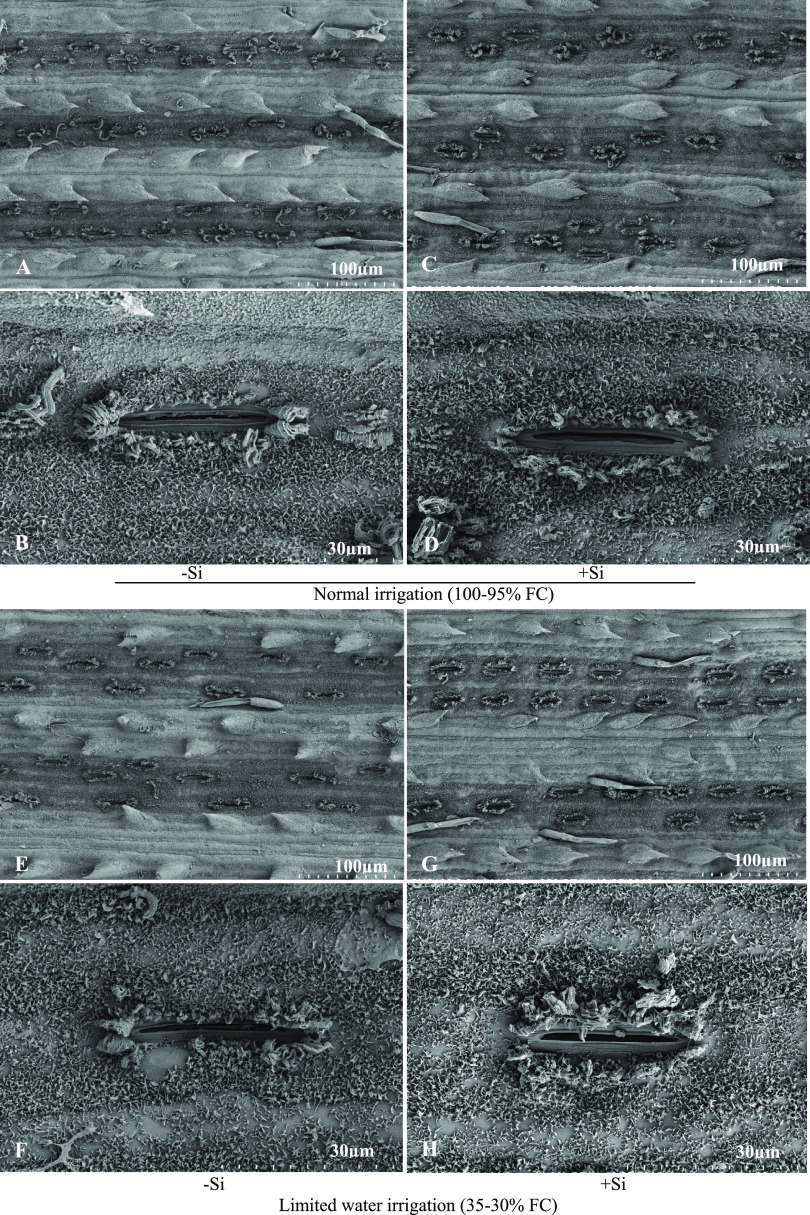
Figure 3.
Scanning electron micrographs of stomata ultrastructure in the epidermis of sugarcane leaves in normal (A, B) and limited water supply treatments without Si (E, F) or with Si supplementation for normal (C, D) and limited water (G, H) at 500 mg L–1 Si. The scale bars for the micrographs were 30 μm (B, D, F, and H) and 100 μm (A, C, E, and G), respectively.

