
Figure 3:
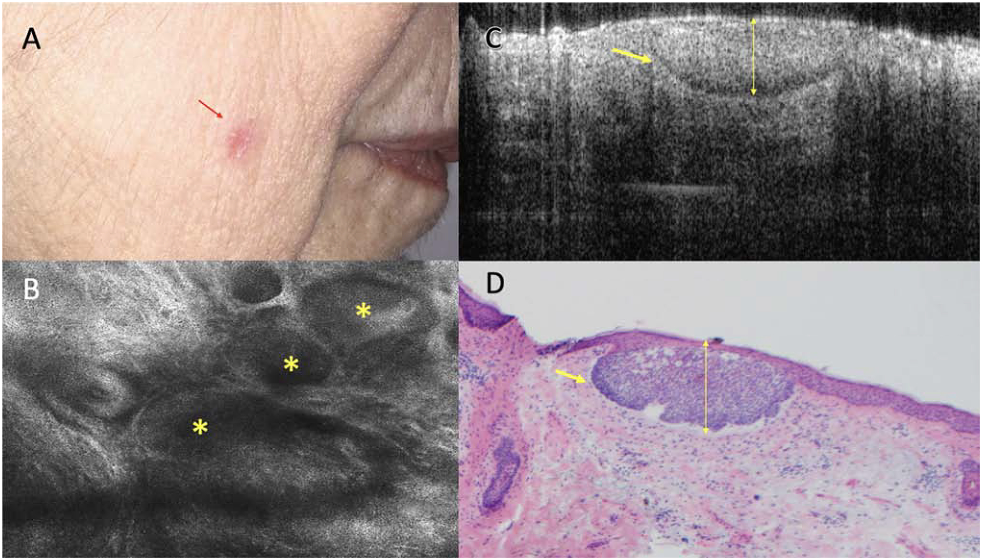
Superficial basal cell carcinoma (BCC) on the cheek with no clinical evidence of residual tumor. A. Clinical appearance of the biopsy site (red arrow). B. Reflectance confocal microscopy showing tumor islands (yellow asterisk) with palisading and clefting (750 × 750 μm). C. Optical coherence tomography image showing hyporeflective tumor island with retraction space (yellow arrow) measuring 200 μm in depth (1000 × 2000 μm) D. Mohs frozen section showing corresponding superficial BCC (yellow arrow), 200 μm depth. (H&E, 4X magnification).