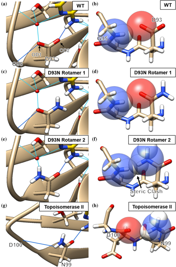
FIGURE 6.

Hsp90α hydrogen bonding network for D93 (a, PDB: 3T10), modeled rotamers for D93N (c and e) and the N99 residue of Topoisomerase II (g, PDB: 4GFH). Water‐mediated bonds are in light blue and direct hydrogen bonds are in dark blue. (b) Hsp90α D93 is sterically allowed. (d) Rotamer 1 of D93N is sterically allowed but cannot act as a hydrogen‐bond acceptor to bound nucleotide or inhibitor. (f) Rotamer 2 is sterically disallowed with an atomic overlap of 0.8 Å. (h) The N99 residue of Topoisomerase II does not sterically clash with the backbone. Rotamer modelling was performed with Chimera
