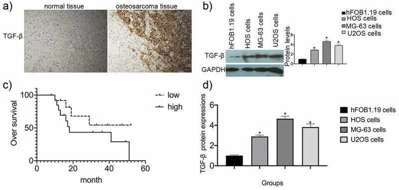
Figure 1.

TGF-β expression in osteosarcoma specimens and cell lines. (a) Immunohistochemical staining was used to analyze TGF-β expression in both osteosarcoma specimens and normal adjacent tissues. (b) Survival analysis was performed using the Kaplan-Meier method. Data are presented as mean ± SEM of three independent experiments. (c) TGF-β protein levels in osteosarcoma cell lines (*P < 0.05). (d) TGF-β mRNA expression in osteosarcoma cell lines (*P < 0.05).
