FIGURE 2.
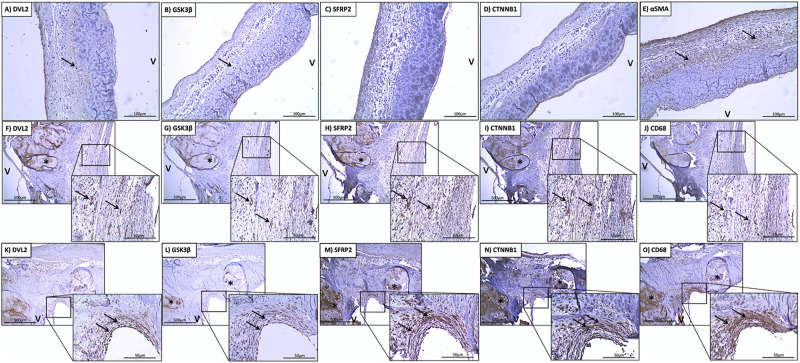
Stenotic aortic valves have greater immunostaining of Wnt/β-catenin mediators compared to healthy valves. Low (A) DVL2, (B) GSK-3β, (C) SFRP2, (D) β-catenin immunoreactivity observed in histologically normal leaflet (arrows indicate weak immunostaining in native cells). Prevalent immunostaining of (E) αSMA in histologically normal leaflets. Co-localization of (F) DVL2, (G) GSK-3β, (H) SFRP2 (I), β-catenin immunoreactivity with macrophages around areas of calcification (arrows), indicated by (J) CD68 staining. Co-localization of (K) DVL2, (L) GSK-3β, (M) SFRP2 (N), β-catenin immunoreactivity in valve interstitial cells in the thickened fibrosa (arrows), indicated by (O) αSMA staining. *Calcified foci; v, ventricular side of the leaflet. Arrows point to regions of interest.

