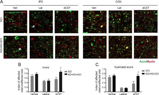
Figure 6.
The effect of ketone salt combined with the ketogenic diet on axonal sparing in spinal cord of spinal cord injury rats.
(A) Representative images of spinal cord tissue 2.5 mm caudal to the epicenter immunostained for axons (β-tubulin III and SMI-312; green) and myelin (MBP; red) 8 weeks post-injury, both ipsilateral (IPS) and contralateral (CON) to injury. Yellow arrows indicate examples of myelinated axons. Gray arrows indicate examples of putative myelin debris. Bar scale: 20 μm. (B, C) Quantification of axons (B) and myelinated axons (C) in the KD + KS + SCI (black; n = 12) and SCI groups (gray; n = 12) 8 weeks post-injury in the dorsal corticospinal tract (dCST), lateral white matter (Lat), and ventral white matter (Ven). Numbers were normalized to the contralateral side (index). Data are expressed as the mean ± SEM (n = 5 per group). †P < 0.05, vs. SCI group (two-tailed unpaired Student’s t-tests). MBP: Myelin basic protein.

