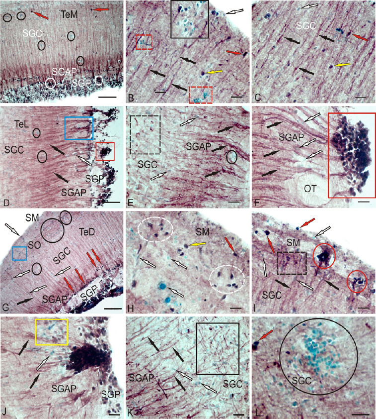
Figure 9.

CBS in the optical tectum of rainbow trout Oncorhynchus mykiss at 1 week after UEI.
(A) In the medial zone of tectum (TeM); CBS+ cells in the SGC are indicated by red arrows; reactive neurogenic niches (RNN) are outlined by black ovals; and CBS+ cells in the SGP are indicated by white ovals. (B) Large RNN in the stratum marginale (SM) (black square), small RNN (red dotted square), radial fibers (by black arrows), CBS– cells (white arrow), intensively labeled cells (red arrow), and moderately labeled cells (yellow arrow). (C) CBS+ radial glia fibers (black arrows) in the SGC (other designations see in В). (D) CBS+ cells forming numerous dense clusters in the SGP (in the red rectangle) and bundles of CBS+ radial fibers in the SGAP (in blue rectangle); radial fibers are indicated by black arrows and CBS– cells are indicated by white arrow). (E) Clusters of activated CBS+ astrocytes (outlined by black dotted line) in the lateral tectum (TeL); reactive neurogenic niches (RNN) are outlined by black oval. (F) CBS– cells (white arrows) located among fibers of the lateral optical tract; radial fibers (by black arrows), red rectangle outlines a cluster of CBS+ cells at a higher magnification; (G) CBS-migrating cells in the SM (in blue rectangle) and RNN in the SO (in black ovals) of the TeD; (H) Diffuse group of CBS+ cells (in white dotted ovals) in the SM at a higher magnification. (I) Dense cluster of CBS+ cells in the SM (in red ovals); CBS+ radial glia in black dotted rectangle. (J) A large single cluster of CBS+ cells, next to CBS-negative RNN (in yellow rectangle). (K) Clusters of tangentially located CBS+ reactive astrocytes (in black rectangle); L – Large parenchymal RNN (in oval) in the SGC. Immunoperoxidase labeling of CBS in combination with methyl green staining. Scale bars: 100 µm in A, D, G and 20 µm in B, C, E, F, H–L. CBS: Cystathionine β-synthase; RNN: reactive neurogenic niches; SGAP: stratum griseum et album periventriculare; SGC: stratum griseum centrale; SGP: stratum griseum periventriculare; SM: stratum marginale; SO: stratum opticum; TeD: dorsal tectum; UEI: unilateral eye injury.
