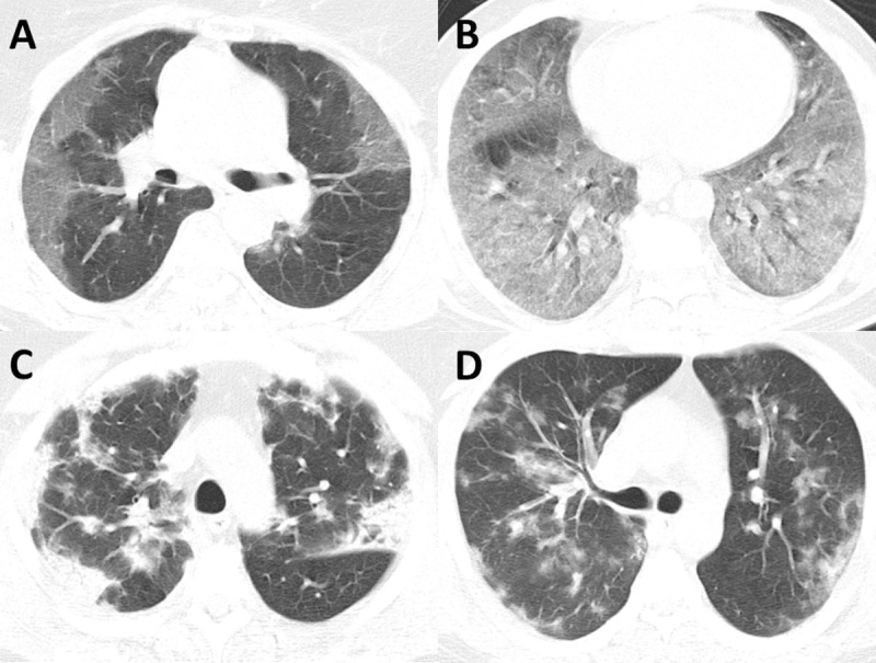
Fig 1. Examples of opacification patterns of COVID-19 on CT imaging.

(A) Most common presentation of COVID-19 on CT imaging with multifocal peripheral ground-glass opacities. (B) Example of diffuse multi-lobar ground-glass opacities in COVID-19. (C) Example of consolidative and ground-glass opacities with both peripheral and central distribution. (D) Example of rare bilateral nodular consolidations seen in COVID-19.
