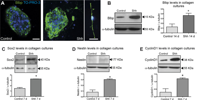
Fig 4. Shh promotes proliferation and maintenance of RGCs.
Differentiation experiments were performed during 14 days without addition of EGF or FGF-2. (A) Representative images of immunostaining for Blbp after control or Shh treatment. Close-up view of a RGC stained for Blbp treated with Shh. Bar, 20 μm. (B) Western blot and densitometry analysis for Blbp expression show higher levels in Shh treated cultures. Western Blot analysis of Sox2 (C), and Nestin (D) levels after 7 days of treatment with Shh indicate an increase of neural progenitors. (E) Western blot of Cyclin D1, a read-out response to Shh pathway activation, indicates an increased proliferation even in absence of other additional growth factors after 7 days of Shh incubation. *, p<0.05. In Fig 4C, lane 1 and 2 of each panel present non-adjacent lanes of the same original blot; additional lanes were removed from the image in preparing the figure. The original image data supporting Fig 4C are in S2 File, and data supporting Fig 4B, D, E are in S1 and S3 Files.

