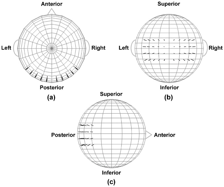
Figure 4.
The 4-shell spherical head forward volume-conduction model (only the outer shell surface is represented in the figure). (a) Top head view; (b) Rear head view; (c) Side head view. Black arrows indicate the locations and orientations of the extrastriate dipole sources. The remainder of the spherical surface was filled with 148 equally-spaced background activity dipoles (not shown). All dipoles were placed at superficial (2mm subdural) cortical locations. Figure adapted from [11] with permission of the authors.

