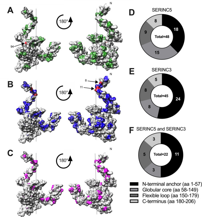
Fig 3. Structural distribution of Nef residues associated with SERINC antagonism.
(A, B, C) The location of Nef polymorphisms associated with internalization of SERINC5 (A, green), SERINC3 (B, blue), or both proteins (C, magenta) are indicated on a composite structural model of HIV Nef (based on PDB 2NEF and 1QA5) (Lamers, PLoS One 2011). Residue 94 (SERINC5, panel A) and residues 8 and 11 (SERINC3, panel B) are highlighted in red. (D, E, F) The distribution of natural polymorphisms associated with internalization of SERINC5 (n = 48) (D), SERINC3 (n = 45) (E) or both proteins (n = 22) (F) among Nef’s major functional domains is illustrated.

