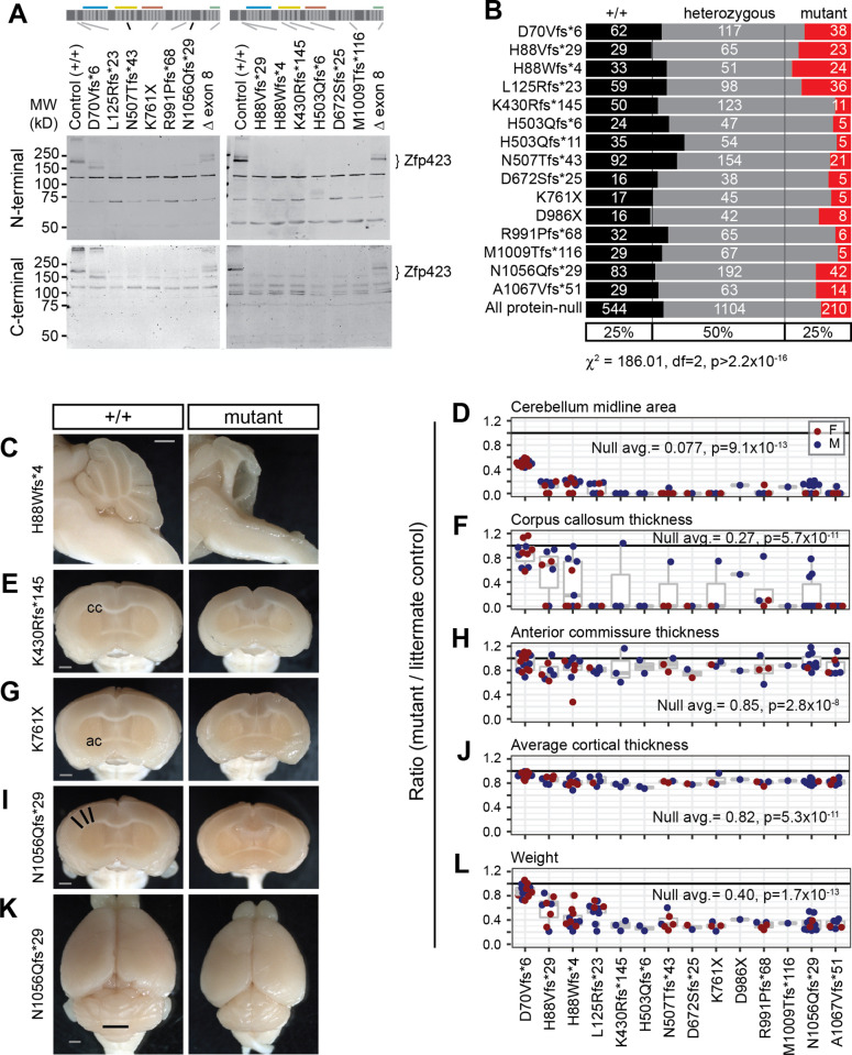
Fig 2. Zfp423 frameshift and nonsense mutations are effectively null, except D70Vfs*6.
(A) Western blots detected full length Zfp423 in perinatal cerebellum of control samples. Among PTC homozygotes, D70Vfs*6 and a deletion of terminal exon 8 showed altered-size proteins at reduced levels. No consistent evidence for residual protein was seen for any other PTC variants, detection threshold ≤5% wild-type level. N-terminal, Bethyl A304-017A. C-terminal, Millipore ABN410. Cross-reacting background bands were independent of PTC position. (B) Reduced frequency of homozygotes for each PTC at biopsy (P10-P20) from breeding records. Summary chi-square is for protein-null alleles (excluding D70Vfs*6). (C) Mid-sagittal images showed variable amount of midline cerebellar tissue in mutants, with residual tissue attributable to hemispheres. (D) Ratio of midline cerebellum area (mutant/control) from block face images. Coronal block face images showed abnormal forebrains, including (E, F) disrupted or reduced corpus callosum (cc), (G, H) reduced anterior commissure (ac), and (I, J) reduced cortical thickness, measured as the average radial distance at 15°, 30° and 45° from midline (black lines). (K) Representative surface views. Vermis width as plotted in Fig 1 is indicated (black line). (L) PTC mutants had reduced body weight at sacrifice. (D, F, H, J, L) Averages and Wilcoxon signed-rank test p-values for ratio = 1 for combined data from all PTC alleles excluding D70Vfs*6 are shown. Female pairs red, male blue. Scale bars, 1 mm.

