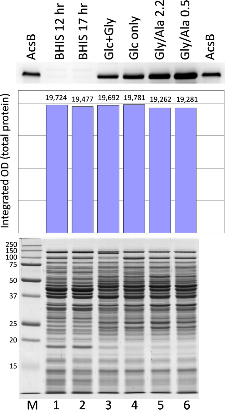
FIG 6.
Western blot analysis of AcsB levels in different growth media. Cell extracts prepared from the large-scale growths indicated were analyzed by SDS-PAGE. Gels containing 12.5 μg total protein per lane were stained with Coomassie blue for protein (bottom, lanes 1 to 6), and 37.5 μg per lane of the same samples was applied for Western blot assays of AcsB (top). Gly/Ala 2.2 and Gly/Ala 0.5 indicate Gly/Ala growths with 2.2 and 0.5 mM glucose remaining at the time of harvest. Purified AcsB, 10 ng, was applied on the outer lanes of the Western blot, and molecular weight markers (M) were included on the stained gel. The estimation of equivalent amounts of protein loaded was verified by densitometric analysis of the stained gel (center).

