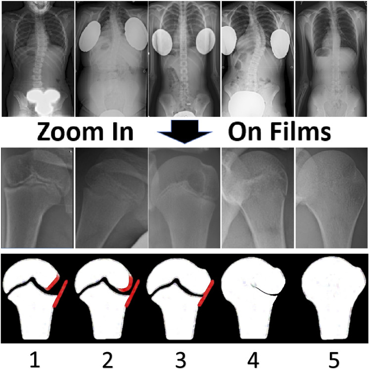
Fig. 1.
Evaluation of humeral head stage on spine radiographs. Representative images from spine radiographs that were collected between 2007 and 2017 are shown in the top row. The zoom function that was built into the Visage visualization software used by our institution was used to magnify the humeral head, as shown in the middle row. Schematic illustrations depicting the relevant features of the humeral head during each stage are shown in the bottom row. In Stage 1, the lateral epiphysis is incompletely ossified, leaving an oblique lateral margin; this results in a radiolucent triangular gap between the epiphyseal and metaphyseal margins. In Stage 2, increased ossification leads to a rounded epiphyseal margin. In Stage 3, the metaphyseal and epiphyseal lateral margins are colinear or capped. In Stage 4, the lateral part of the physis thins and begins partial fusion starting medially; however, the fusion is not complete and the most lateral portion remains open. Finally, Stage 5 is analogous to Risser 5, with complete fusion.

