
Figure 4.
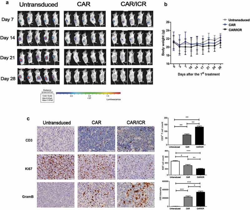
PSMA-CAR-T cells co-expressing ICR significantly enhanced the anti-tumor effects in the xenograft model.
a. In vivo image of LAPC-9-luc xenograft model. b. Quantitative value of fluorescence intensity of tumors in vivo in each treatment group. c. Variations in the body weight of the xenograft model inoculated with tumor cells and after treatment (n = 5, bar value represents the dispersion degree). d. Immunohistochemical expression of CD3, Ki67, and granzyme B in tumor cells. Each datum represents three independent experiments (n = 3, bar value represent the dispersion degree, ns not significant, p > .05, * p < .05, ** p < .01, *** p < .001).