
Figure 5.
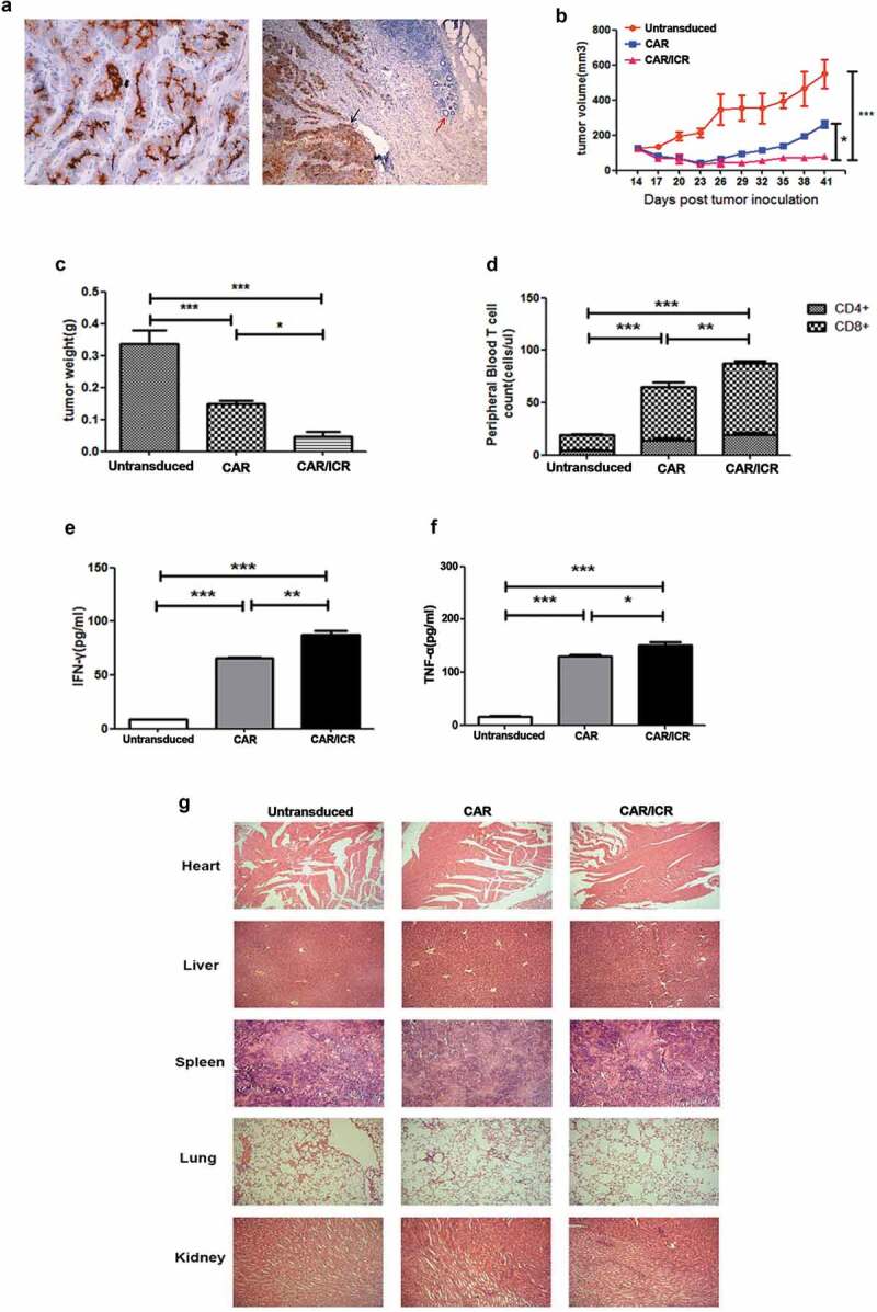
PSMA-CAR-T cells co-expressing ICR significantly enhanced the anti-tumor effects in the PDX model.
a. Immunohistochemical analysis of PSMA antigen expression in patient-derived tissues. b. After the establishment of the PDX model, CAR-T cells were administered through the caudal vein for the experiment groups and untransduced T cells for the control group. Tumor volume was measured with a vernier calliper every 3 days. c. On the 41st day, mice were sacrificed and tumor weight was measured. d. 100 μL of peripheral blood of the mice was collected on the 7th day after the infusion of effector cells, and the number of CD4+ and CD8+ T cells was counted. e. Level of IFN-γ in the peripheral blood of mice. f. Levels of TNF-α in the peripheral blood of mice. Each datum represents 3 independent experiments (n = 6, bar value represent the dispersion degree, * p < .05, ** p < .01, *** p < .001). g. Safety analysis of major organs after CAR-T cell treatment. The hearts, livers, spleens, lungs, and kidneys of each group were embedded in paraffin and cut into sections, which were stained with hematoxylin and eosin. Images were taken at 10 × magnification.