Figure 1.

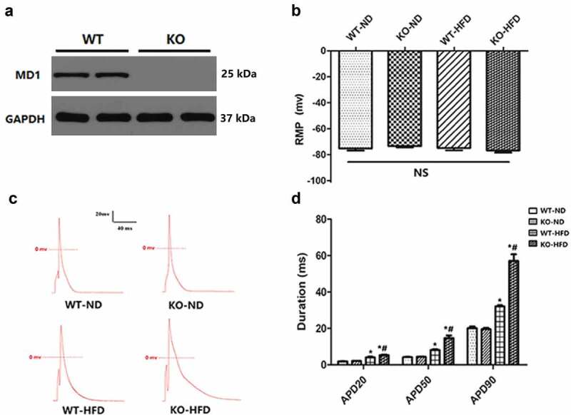
MD1 Deletion alters action potential durations in ventricular cardiomyocytes isolated from WT and KO mouse hearts after 20 weeks of ND or HFD feeding. (a) Representative western blots of MD1 expression in LV tissues from WT and MD1-KO mice (n = 6). (b) Changes of resting membrane potential (RMP) (n = 8 cardiomyocytes from n = 4 mice each group). (c, d) Representative action potential figures and statistical analysis of the 20%, 50%, and 90% action potential durations (n = 8 cardiomyocytes from n = 4 mice each group). Data are expressed as mean ± SEM. * p < 0.05 vs. WT-ND group, # p < 0.05 vs. WT-HFD group.
