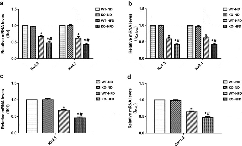
Figure 4.

Expression of channels in WT and KO mouse hearts after 20 weeks of ND or HFD feeding. (a-c) The mRNA levels of the potassium channel subunits of transient outward potassium current Ito,f (Kv4.2, Kv4.3), delayed rectifier potassium current IK, slow (Kv1.5 and Kv2.1), and inwardly rectified potassium current IK1 (Kir2.1). (d) The mRNA levels of the calcium channel subunit of L-type calcium current ICaL (Cav1.2). N = 4 mice in each group. Data are expressed as mean ± SEM. * p < 0.05 vs. WT-ND group, # p < 0.05 vs. WT-HFD group.
