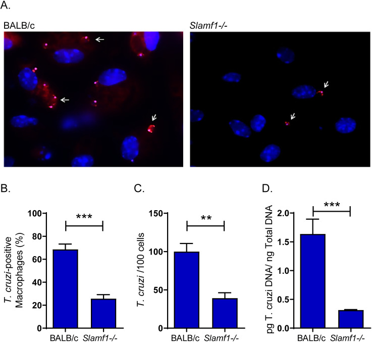
Fig 1. Interaction of T. cruzi with macrophages from BALB/c and Slamf1-/- mice.
A) Fluorescence microscopy images corresponding to BALB/c and Slamf1-/- macrophages incubated for 1 h with parasites of the Y strain. The nucleus of macrophages appears stained in blue (Dapi). The arrows point to fluorescent parasites labelled with Mito Tracker Orange attached to macrophages from BALB/c and Slamf1-/- mice. A representative experiment of two is shown. B) Percentage of peritoneal macrophages from BALB/c and Slamf1-/- mice interacting with T. cruzi after 1 h. C) Number of trypomastigotes attached to 100 peritoneal macrophages from BALB/c and Slamf1-/-mice after 1 h. In B and C the mean ± SEM corresponds to 14 fields of each condition performed in duplicate. D) Quantification of the parasite load in macrophages from BALB/c and Slamf1-/- mice incubated with the Y strain during 1h. The statistical significance of the differences between both strains of mice is indicated: *** p <0.001, ** p <0.01.

