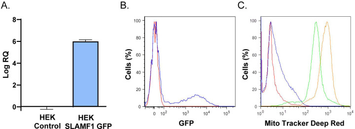
Fig 2. Interaction of T. cruzi with HEK293T cells over-expressing SLAMF1-GFP.
A) Quantification by RTqPCR of the relative expression of SLAMF1-GFP mRNA respect to human 18S mRNA in HEK Control and HEK SLAMF1 GFP cells. B) Flow cytometry analysis of SLAMF1 Protein expression in HEK Control cells (Red line) and HEK SLAMF1 GFP cells (blue line). C) Flow cytometry analysis of HEK Control cells (green line) and HEK SLAMF1 GFP cells (orange line) incubated with T. cruzi trypomastigotes labelled with Mito Tracker Deep Red); HEK Control cells (Red line) and HEK SLAMF1 GFP cells (blue line) were not incubated with labelled parasites. A representative result out of two independent experiments is represented.

