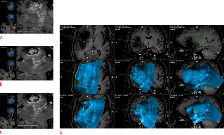
Fig. 12. Resection control scans using navigated two-dimensional ultrasonography (2DUS) versus navigated three-dimensional ultrasonography (3DUS).
A-C. The left column depicts serially-acquired navigated 2DUS images. Note the differing planes of each 2DUS image as shown in the corresponding magnetic resonance cuts. D. In the same patient, after acquiring a 3DUS scan, serial scans can be depicted side-by-side and visualized in the same planes, allowing for easier comparison.

