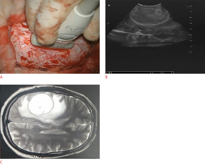
Fig. 5. Two-dimensional ultrasonography.
A. Probe is positioned over the surface of the dura. B. Two-dimensional ultrasound image obtained in axial plane is depicted. C. Corresponding magnetic resonance image is seen in this image. It may not always be feasible to obtain such true anatomical planes, depending on the craniotomy aperture and location.

