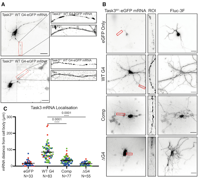
Figure 4.
Task3 5′ UTR (GGN)13 repeat is required for distal neurite localisation of Task3 mRNA. (A) RNA FISH of DIV10 embryonic murine cortical neurons transfected with WT G4 Task3M1-eGFP. Cells were hybridised with anti-eGFP probes and anti-MAP2/Tau primary antibodies to determine neurite localisation of exogenous Task3 mRNA. Image 2 was taken at two exposures to define mRNA granules in both the cell body and neurites with the respective images merged (dotted line). Original un-edited images available in Supplementary Figure S5. (B) RNA FISH of transfected DIV10 embryonic murine cortical neurons co-transfected with mutant G4 5′ UTR Task3M1-eGFP and Fluc-3F as a soluble whole-cell marker. Cells were hybridised with anti-eGFP probes and anti-FLAG M2 primary antibody showing localisation of exogenous mutant G4 5′ UTR Task3M1-eGFP mRNA in comparison to whole-cell staining. (C) The distance of exogenous mutant G4 5′ UTR Task3M1-eGFP mRNA from the cell body, quantified in Fiji for cells possessing neurites ≥100 μm in length. Number of neurites analysed is shown from three blinded biological replicates (coloured groups), where error bars represent SEM. Statistical analyses were carried out by one-way ANOVA (F(3, 247) = 89.01, P < 0.0001), with post-hoc Sidak's multiple comparison tests, P values shown. Scale bar = 20 μm.

